UCLA paleobiologist J. William Schopf and colleagues have produced 3-D images of ancientfossils — 650 million to 850 million years old — preserved in rocks, anachievement that has never been done before.
If a future space mission to Mars brings rocks back toEarth, Schopf said the techniques he has used, calledconfocal laser scanning microscopy and Ramanspectroscopy, could enable scientists to look at microscopic fossils inside therocks to search for signs of life, such as organic cell walls. These techniqueswould not destroy the rocks.
"It's astounding to see an organically preserved,microscopic fossil inside a rock and see these microscopic fossils in threedimensions," said Schopf, who is also a geologist,microbiologist and organic geochemist. "It's very difficult to get any insightabout the biochemistry of organisms that lived nearly a billion years ago, andthis (confocal microscopy and Raman spectroscopy)gives it to you. You see the cells in the confocalmicroscopy, and the Raman spectroscopy gives you the chemistry.
"We can look underneath the fossil, see it from the top,from the sides, and rotate it around; we couldn't do that with any othertechnique, but now we can, because of confocal laserscanning microscopy. In addition, even though the fossils are exceedingly tiny,the images are sharp and crisp. So, we can see how the fossils have degradedover millions of years, and learn what are real biologicalfeatures and what has been changed over time."
His research is published in the January issue of thejournal Astrobiology, in which he reports confocalmicroscopy results of the ancient fossils. (He published ancient Ramanspectroscopy 3-D images of ancient fossils in 2005 in the journal Geobiology.)
Since his first year as a Harvard graduate student in the1960s, Schopf had the goal of conducting chemicalanalysis of an individual microscopic fossil inside a rock, but had notechnique to do so, until now.
"I have wanted to do this for 40 years, but there wasn't anyway to do so before," said Schopf, the firstscientist to use confocal microscopy to study fossilsembedded in such ancient rocks. He is director of UCLA's
Raman spectroscopy, a technique used primarily by chemists,allows you to see the molecular and chemical structure of ancient microorganismsin three dimensions, revealing what the fossils are made of without destroyingthe samples. Raman spectroscopy can help prove whether fossils are biological, Schopf said. This technique involves a laser from amicroscope focused on a sample; most of the laser light is scattered, but asmall part gets absorbed by the fossil.
Schopf is the first scientist touse this technique to analyze ancient microscopic fossils. He discovered thatthe composition of the fossils changed; nitrogen, oxygen and sulfur wereremoved, leaving carbon and hydrogen.
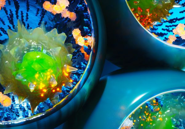
Confocal microscopy uses a focusedlaser beam to make the organic walls of the fossils fluoresce, allowing them tobe viewed in three dimensions. The technique, first used by biologists to studythe inner workings of living cells, is new to geology.
The ancient microorganisms are "pond scum," among theearliest life, much too small to be seen with the naked eye.
Schopf's UCLA co-authors includegeology graduate students Abhishek Tripathi and Andrew Czaja, andsenior scientist Anatoliy Kudryavtsev.The research is funded by NASA.
Schopf is editor of "Earth'sEarliest Biosphere" and "The Proterozoic Biosphere: AMultidisciplinary Study," companion books that provide the most comprehensiveknowledge of more than 4 billion years of the earth's history, from theformation of the solar system 4.6 billion years ago to events half‑a‑billionyears ago.
-UCLA-
SW051