Key takeaways
- UCLA bioengineers have improved a research technology they developed — tiny bowl-shaped particles that trap one to a few cells and their secretions for analysis — by adding a cap to the compartments.
- A new study shows that capping the particles, called nanovials, enables experiments with additional cell types, with growing colonies and with interactions between cells.
- Capped nanovials could benefit drug discovery, basic biological studies, chemical manufacturing and artificial intelligence–aided research.
A UCLA-developed technology that is like a test tube but on the cellular scale has led to a number of biomedical discoveries in recent years, including revelations about cancer, heart disease and the genetic levers that push cells to release proteins.
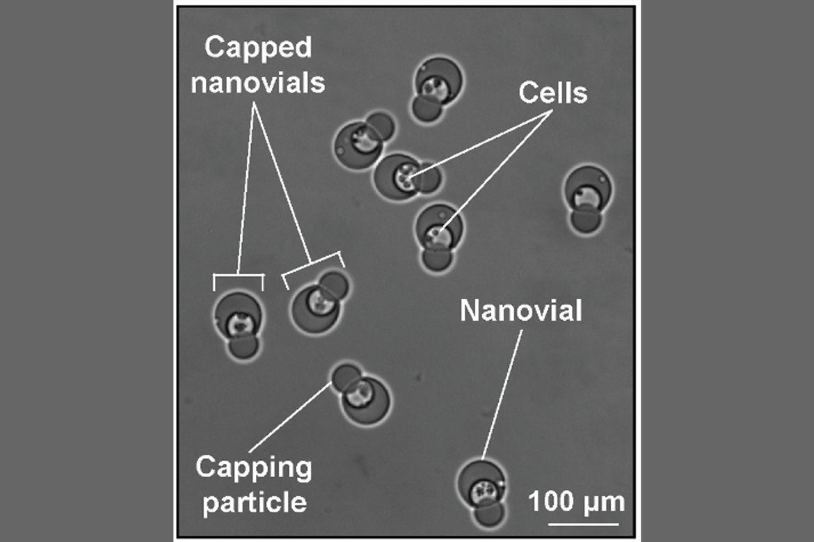
Imagine shrinking a test tube until it can hold just one or two cells, then making hundreds of thousands of these miniature test tubes to run experiments simultaneously. That’s the idea behind UCLA’s nanovial technology, which uses tiny bowl-shaped hydrogel particles to capture cells and measure what they produce. Now, researchers have added a lid, allowing cells to remain inside longer, grow into small colonies and interact with other cells under controlled conditions.
A new study published in Science Advances demonstrated that capped nanovials can be used to collect and sort cells, including bacteria and yeast that were difficult to capture in uncapped nanovials. By confining cells for longer, the capped nanovials enabled researchers to grow cells inside of them and select for the fastest-growing colonies. Profiling interactions between cells, the team used capped nanovials to confirm that one cell changed the behavior of another.
“On their own, nanovials compartmentalize cells and their products, but it’s kind of short-term,” said corresponding author Dino Di Carlo, the Armond and Elena Hairapetian Professor of Engineering and Medicine and chair of the bioengineering department at the UCLA Samueli School of Engineering. “The capping system opens up other types of longer-term experiments that involve growing colonies or selecting bacteria or yeast based on certain characteristics.”
Turning a glitch into an enhancement
The idea of capping nanovials began with a quirk in the technology’s previous generation. The researchers observed that one nanovial would occasionally nest in the cavity of another, blocking the bottom one from doing its job.
“You never know where ideas are going to come from,” said Di Carlo, a member of the California NanoSystems Institute at UCLA. “At first we said, ‘Oh, that’s a problem,’ but then we asked, ‘What if we turn it into a solution?’”
The caps are tiny hydrogel spheres. To help link the bowl and cap, the investigators decorated them with two molecules that bond together like molecular Velcro: vitamin B7 around the rim of the nanovial and a certain bacterial protein around the cap.
The actual connection happens through a seemingly random process. Caps and nanovials were spun in the same solution using a centrifuge, then allowed to settle. Nanovials landed first with their cavities facing up, then caps fell into place.
After the researchers optimized the self-assembly process, as many as nine out of 10 nanovials ended up capped. Importantly, because both the nanovial and cap are porous, nutrients and waste pass through freely while cells stay trapped inside.
New capabilities
Unlike many miniature cell-testing systems that require specialized instruments, capped nanovials can be analyzed and sorted using equipment already found in many biological research laboratories. This could make sophisticated experiments accessible to a much wider range of scientists.
Uncapped nanovials are effective in corralling mammalian cells. This is due to coatings on their interior that bind to proteins on a particular cell’s surface. However, bacteria and yeast generally lack those proteins and so they tend to drift out. The study showed that these types of cells remained confined inside capped nanovials even though they weren’t attached.
“We don’t need to find a particular way to capture each species, and that lowers the barrier for someone to apply this approach,” said Di Carlo, a cofounder of Partillion Bioscience, which commercializes nanovials. “The caps move nanovials toward being a generalized platform that doesn’t require tuning, the same way you can use a petri dish to look at any type of cell.”
When cells divide inside uncapped nanovials, the new cells usually spill out. The researchers found that capped nanovials supported the growth of small colonies of bacteria for six hours, yeast for 12 hours and engineered mouse cells for three days before growth led cells to pop the lid.
While encapsulated, the minicolonies could be sorted by their growth rate. The caps also improved secretion measurements for cells that release especially large amounts of protein, where the extra containment gave a cleaner signal than uncapped nanovials.
Possibilities for impact
Being able to identify and recover cells that grow quickly under less-than-optimal manufacturing conditions, or that produce especially useful molecules, could boost applications in which cells serve as tiny factories. Examples span a range of industries from the manufacturing of pharmaceuticals and protein medicines such as insulin to the brewing of beer.
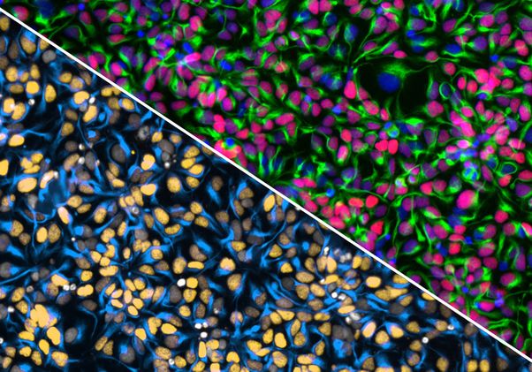
The researchers also demonstrated that capped nanovials can serve as miniature laboratories for studying how cells communicate. They placed antibody-producing cells together with engineered T cells that glow green when activated by the antibody.
By keeping the cells and their signals together, the capped nanovials made it easier to identify which antibody-producing cells activated the T cells — a capability that could help researchers discover more effective therapeutic antibodies. Rather than simply measuring how much of a molecule a cell produces, researchers could now test whether that molecule actually changes the behavior of another cell.
“We’ve showed proof of principle for functional drug discovery tests,” Di Carlo said. “You could also imagine basic studies putting any two cells together and looking at how they change each other over time through contact and secretion.”
For next steps, the investigators are exploring scientific applications such as screening bacteria for antibiotic susceptibility. They’re also evaluating the flexibility of capped nanovials for more complex experiments that involve changing the conditions of captured cells over time.
With the expanded palette of capabilities — and the opportunity to conduct thousands to millions of cell- and colony-scale experiments in parallel — the capped nanovial may have special advantages for biomedical science in the age of AI. Artificial intelligence could eventually help scientists predict which cells will grow fastest, produce useful medicines or respond to specific treatments. But developing such models requires enormous amounts of experimental data.
“These are very data-hungry systems, and it takes huge, high-quality datasets to create generalizable models,” Di Carlo said. “We can’t do five experiments in a day anymore. We have to do 100,000 experiments in a day.”
Authors and funding
The study’s co-first authors are Michael Mellody, who earned both his bachelor’s and Ph.D. in bioengineering from UCLA Samueli, and UCLA Samueli postdoctoral researcher Yuta Nakagawa. Other co-authors are affiliated with UCLA, Partillion Bioscience and the University of Washington.
This research was funded by the National Institutes of Health, the Chan Zuckerberg Initiative, the Japan Society for the Promotion of Science and the Nakatani Foundation.